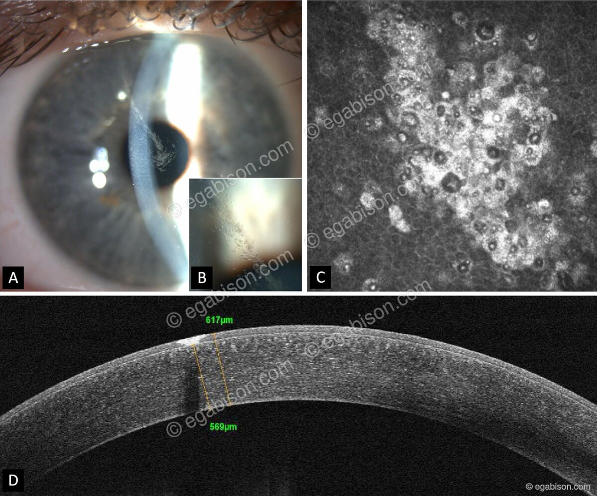
A 20-year-old man, with no family history, is referred by his ophthalmologist for a unilateral corneal lesion. What diagnosis would you consider?
A & B: slit lamp. C: in vivo confocal microscopy. D: anterior-segment OCT.
Lisch epithelial corneal dystrophy (X-linked)
A band-shaped, whorled microcystic epithelial infiltrate extending from the limbus toward the visual axis. In vivo confocal microscopy shows hyperreflective cells and hyporeflective cystic spaces within the epithelium; anterior-segment OCT confirms the epithelial location with an intact Bowman's layer.
The trap: this dystrophy is frequently mistaken for herpetic keratitis (dendritic ulcer) and wrongly treated with aciclovir.
Reference: Martin G-C, Guindolet D, Gabison E. Lisch epithelial corneal dystrophy: a misdiagnosed clinical entity? J Fr Ophtalmol. 2024;47:104245.