2.Les paupières et le bord libre
L'examen du bord libre palpébral est systématique : télangiectasies, épaississement, irrégularité ou déplacement des orifices meibomiens. Le test à la pression apprécie la qualité du meibum. Un meibum épais, opaque ou absent signe un dysfonctionnement des glandes de Meibomius (DGM), première cause de sécheresse évaporative.
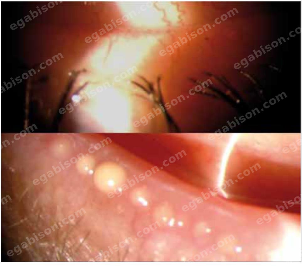
L'apport majeur depuis 2011 est la meibographie infrarouge non contact, qui visualise l'architecture des glandes et quantifie leur perte (cotée de 0 à 4 selon le pourcentage de surface tarsienne dépourvue de glandes).
3.Le film lacrymal
Le film lacrymal se juge sur sa stabilité (BUT/NIBUT), son volume (ménisque lacrymal, mesurable en OCT du segment antérieur) et sa composition. Deux marqueurs ont structuré la pratique moderne : l'osmolarité lacrymale et la MMP-9 (marqueur inflammatoire de surface).
2.The eyelids and lid margin
Examination of the lid margin is systematic: telangiectasia, thickening, irregularity or displacement of the meibomian orifices. The expression test assesses meibum quality. Thick, opaque or absent meibum indicates meibomian gland dysfunction (MGD), the leading cause of evaporative dry eye.
The major advance since 2011 is non-contact infrared meibography, which images gland architecture and quantifies gland loss (graded 0 to 4 according to the percentage of tarsal area devoid of glands).
3.The tear film
The tear film is judged on its stability (BUT/NIBUT), its volume (tear meniscus, measurable by anterior-segment OCT) and its composition. Two markers have shaped modern practice: tear osmolarity and MMP-9 (a surface inflammatory marker).